
Contents
Skin staples have become an increasingly common wound closure method in veterinary medicine, used alongside traditional sutures and topical adhesives as part of a comprehensive surgical toolkit. For practitioners who have not used them extensively, or for those looking to understand the clinical reasoning behind their selection, a thorough answer to the question of how do skin staples work provides the foundation for confident and appropriate use.
This article explains the mechanics of skin staple application, examines the clinical advantages that lead veterinary professionals to choose a disposable skin stapler over conventional sutures in specific situations, and outlines the important considerations around patient selection, technique, and post-operative care.
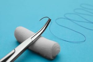
A skin staple is a small, precisely formed piece of stainless steel or titanium wire shaped into a rectangular configuration. Medical staples used in veterinary and human skin closure are designed to a specific geometry that determines how they engage tissue, how much pressure they apply to wound edges, and how they are removed after healing.
The cross-section of a surgical skin staple is not a flat strip of metal. It has a slightly curved or rectangular profile that, when formed correctly against the skin surface, creates what is known as a formed staple shape that provides secure tissue engagement without cutting through or crushing the skin beneath it.
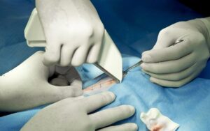
A medical staple gun, also called a skin stapler or surgical stapler, is a handheld device that contains a cartridge of pre-loaded staples. When the practitioner squeezes the handle or trigger of the device, a mechanism within the stapler drives a single staple downward through a forming plate, bending the legs of the staple around and partially under the tissue to create the closed staple configuration.
When the device is positioned against the skin surface with wound edges held in apposition, activating the stapler drives the straight-legged staple downward. The legs of the staple penetrate the skin on either side of the wound line. As the staple descends, the forming plate within the medical stapler bends the legs inward and partially under the tissue, creating the characteristic closed rectangular shape that secures the staple in position.
This forming process happens in a fraction of a second and does not require the practitioner to make any adjustment between staple placement and completion. The device advances automatically to the next staple after each firing, allowing rapid sequential placement along the wound.
Once formed, the closed staple sits against the skin surface with its legs embedded in the tissue on either side of the wound. The bridge of the staple spans the incision line, holding the wound edges together through direct mechanical apposition. The geometry of the formed staple is designed to provide even pressure across the wound edge without excessive compression, which would compromise local circulation and healing.
A well-placed staple should produce slight eversion of the wound edges, meaning the edges turn outward very slightly rather than inverting. Edge eversion is associated with better cosmetic outcomes and more reliable primary intention healing because it ensures the dermal layers on either side of the wound are in direct contact, supporting efficient cellular bridging across the incision.
The disposable skin stapler used in veterinary practice is a single-use device supplied in sterile packaging with a fixed number of staples per cartridge. Common configurations contain between 25 and 35 staples per device. Once all staples have been deployed, the device is discarded. This single-use design eliminates the need for reprocessing, removes the risk of cross-contamination between patients, and ensures consistent performance because the forming mechanism has not been subjected to previous use.
For an overview of the clinical benefits of sterile single-use skin staplers in veterinary practice,the benefits of using sterile disposable skin staplers in veterinary practice provides detailed context.
Modern disposable skin staplers are ergonomically designed to allow consistent staple placement with minimal effort. The device typically features a window or indicator that allows the practitioner to center the staple placement guide over the wound line before firing, ensuring each staple is positioned symmetrically across the incision. Some designs include a staple count indicator that shows how many staples remain in the cartridge.
The handle mechanism is designed to require only a moderate grip force to fire, reducing hand fatigue during longer closures and supporting consistent placement even toward the end of a procedure.
Surgical skin staples are manufactured from stainless steel or titanium. Both materials are biocompatible and resist corrosion in the wound environment. Titanium staples are lighter and may cause less interference with post-operative imaging, which can be a consideration in patients requiring radiographic or MRI assessment of structures near the stapled closure site.
Understanding how do skin staples work in terms of mechanics is only part of the clinical picture. The more practical question for veterinary professionals is when and why a medical stapler represents a better choice than traditional sutures.
The most consistently cited advantage of skin staples over sutures is the speed of application. Each staple is placed in a single trigger action without the need for needle driving, suture manipulation, or knot tying. A closure that would require ten minutes with hand-placed interrupted sutures can often be completed in under two minutes with a disposable skin stapler.
This speed advantage is clinically meaningful in several specific situations. In emergency procedures where patient stability is a concern, rapid skin closure reduces total anaesthetic time and allows the team to shift attention to recovery management. In high-volume practices where multiple procedures are performed daily, the cumulative time saving across cases has genuine operational value. For long incisions such as those associated with orthopaedic approaches or exploratory laparotomy, stapling reduces the fatigue associated with extended hand suturing.
A well-designed disposable skin stapler produces consistent wound edge eversion with each staple, regardless of the experience level of the practitioner. In hand suturing, achieving consistent eversion across the full length of an incision requires deliberate technique and attention to suture depth and spacing. With a skin stapler, the device geometry largely determines the eversion outcome, reducing technique variability.
Consistent wound edge eversion supports reliable primary intention healing and tends to produce a finer, more uniform scar line compared to closures where some sutures have been placed with slightly inverted or uneven tension.
For animals that are more sensitive to prolonged procedures or for patients with borderline cardiovascular or respiratory reserve, minimising the duration of anaesthesia is an important safety consideration. The speed of staple application contributes directly to shorter total procedural time, which benefits at-risk patients.
Stainless steel and titanium are highly biocompatible materials that provoke minimal tissue reaction. The absence of suture material within the tissue layers of the wound reduces the foreign body burden compared to some suture closures. Staples sit on the skin surface rather than passing through multiple tissue layers, which further limits tissue-suture interaction.
Factor |
Skin Staples |
Traditional Sutures |
| Application speed | Very fast, single action per staple | Slower, requires needle and knot technique |
| Technique consistency | High, device-determined | Variable, technique-dependent |
| Wound edge eversion | Consistent | Depends on suture placement technique |
| Tissue reactivity | Very low (metal) | Low to moderate depending on material |
| Removal requirement | Yes, staple remover required | Yes for non-absorbable; no for absorbable |
| Suitable for curved incisions | Less ideal | More flexible |
| Cost per closure | Generally lower per unit | Variable by material |
| Use in contaminated wounds | Acceptable for skin surface | Monofilament preferred in deep layers |
Skin staplers are particularly well suited to long, straight or gently curved incisions. Orthopaedic approaches, lateral thoracotomy incisions, and long abdominal skin closures are all appropriate candidates for staple closure. The speed advantage is most pronounced in these cases, and the consistent staple spacing produces a uniform closure along the full incision length.
In emergency presentations where the priority is rapid, secure wound closure with minimal procedural time, a disposable skin stapler is often the most appropriate external closure tool. The device allows swift skin apposition without the setup time associated with suture material selection and needle handling.
In practices performing high volumes of routine surgeries such as spays, neuters, and minor mass removals, integrating skin staples into the closure protocol can meaningfully reduce per-procedure time. This efficiency benefit accumulates significantly across a full surgical day.
Skin staples are commonly used as the external skin layer following internal closure with appropriate absorbable sutures. After subcutaneous and subcuticular layers have been managed with absorbable materials, staples provide rapid and reliable external apposition. This combination approach is discussed in how disposable skin stapler use supports faster healing for pets.
Skin staples are most effective on straight or gently curved incision lines. In wounds with significant curvature, irregular edges, or complex geometric configurations, traditional sutures offer greater flexibility in placement and can be adapted to the specific shape of the wound more precisely than a stapling device allows.
In very small patients including cats, toy breed dogs, rabbits, and avian patients, the standard dimensions of commercially available skin staples may be disproportionate to the tissue being closed. Fine-gauge sutures allow much more precise tension control in small patients, and the risk of applying excessive pressure with a standard staple size is a relevant concern. Some manufacturers produce smaller staple configurations for paediatric or fine-tissue applications, but availability varies.
In highly visible areas, around the face, or in cases where owners have significant cosmetic expectations, the precision available with fine monofilament sutures or subcuticular absorbable patterns often produces superior results compared to staples. Sutures allow individual tension adjustment at each placement point that staples cannot match.
In animals that are likely to traumatize their wound site, staples present hard metal edges at the skin surface that may cause more local irritation than buried suture material when licked or scratched. In these patients, a subcuticular absorbable pattern combined with tissue adhesive removes external hardware entirely, reducing the target for self-trauma. The role of topical adhesive in these cases is discussed in key benefits of vet skin glue for faster healing in pets.
Skin staples require removal once adequate wound healing has occurred, typically at 10 to 14 days post-operatively in most veterinary skin closures. Removal is performed using a dedicated staple remover instrument, which is designed to invert the formed bridge of the staple, disengaging the legs from the tissue without pulling or tearing.
Correct staple removal technique is important for patient comfort and for avoiding wound disruption. The remover is positioned beneath the staple bridge, and the handles are compressed to invert the staple geometry, allowing clean withdrawal. Attempting to remove staples without the correct instrument risks tearing the tissue and causing unnecessary discomfort.
Detailed guidance on safe and effective staple removal in veterinary patients is provided in practices to remove surgical staples from animal wounds.
The sterility of a disposable skin stapler is maintained through validated packaging that must be intact at the time of use. Before opening, each device should be inspected for packaging damage, moisture, or compromised seals. Any device with suspect packaging should be discarded.
Sourcing disposable skin staplers from reputable veterinary surgical suppliers ensures consistent staple forming quality, reliable firing mechanisms, and guaranteed sterility. Inconsistently manufactured staplers may produce variable staple forming, which affects wound edge eversion and the security of the closure. The role of supply quality in patient outcomes and clinical trust is discussed in the importance of medical supplies in building trust.
Understanding how do skin staples work at a mechanical level, and knowing the clinical reasoning that supports choosing a medical stapler over traditional sutures in specific situations, allows veterinary professionals to integrate this tool effectively into their surgical practice. Skin staples are not a universal replacement for sutures but offer genuine advantages in speed, consistency, and patient handling time that make them the superior choice in long linear closures, emergency cases, and high-volume elective procedures.
Used alongside conventional sutures, absorbable subcuticular patterns, and topical adhesives, the disposable skin stapler is a valuable and practical component of comprehensive veterinary wound closure management.
Strouden supplies high-quality disposable skin staplers and a full range of veterinary wound closure products to support efficient, reliable surgical outcomes. To explore our product range or discuss the right closure solutions for your practice, please contact us today.
A: A medical stapler drives a metal staple through the skin on either side of the wound, then bends the legs inward to form a closed shape that holds wound edges together. Each staple provides mechanical apposition across the incision without suture material or knot tying.
A: A disposable skin stapler is a single-use handheld device pre-loaded with surgical staples. The practitioner positions it over the wound line and fires it to place individual staples in rapid sequence, producing consistent wound edge closure significantly faster than hand-placed sutures.
A: Skin staplers are preferred for long linear incisions, emergency closures where speed matters, and high-volume elective procedures. They are less suitable for curved wounds, very small patients, or areas requiring fine cosmetic precision where sutures offer better control.
A: Yes, skin staples are used in cats and small breed dogs, though practitioners should consider patient size carefully. Standard staple dimensions may be disproportionate in very small patients. In tiny or exotic animals, fine-gauge sutures generally provide better tension control and tissue compatibility.
A: Staples are removed at 10 to 14 days post-operatively using a dedicated staple remover instrument that inverts the staple bridge to disengage the legs cleanly from tissue. Correct technique ensures patient comfort and avoids wound disruption during the removal process.