
Contents
One of the most frequently asked questions among veterinary professionals encountering polydioxanone sutures for the first time is: are PDS sutures dissolvable? The straightforward answer is yes. PDS is an absorbable suture material that degrades within the body through a process called hydrolysis and is eventually eliminated without the need for surgical removal. However, the full clinical picture is considerably more nuanced than this simple confirmation suggests.
PDS suture material has a longer absorption timeline than most other commonly used absorbable sutures, and its degradation can be influenced by patient and environmental factors in ways that occasionally produce unexpected clinical findings. Understanding the complete absorption behavior of PDS, recognizing the signs that suggest degradation is not proceeding normally, and knowing how to respond when those signs appear are all part of using this important suture material competently in veterinary practice.
PDS is the trade designation for polydioxanone, a synthetic absorbable suture produced from a polyester polymer. It is manufactured as a monofilament, meaning the suture consists of a single continuous strand rather than multiple braided filaments. This monofilament construction gives PDS suture material its characteristic smooth surface, which passes through tissue with low friction and resists bacterial wicking along the suture line.
Polydioxanone is a member of the synthetic absorbable suture family, which also includes polyglactin 910, polyglycolic acid, and poliglecaprone 25. Unlike natural absorbable sutures such as chromic catgut, which are broken down by the body’s enzymes, PDS suture material degrades through hydrolysis. Hydrolysis is a chemical reaction in which water molecules progressively cleave the polymer chains that make up the suture, breaking the material down into smaller and smaller fragments until it is fully absorbed and metabolized.
The answer to whether pds suture is absorbable lies in this hydrolytic degradation mechanism. Because PDS undergoes complete breakdown and elimination within the body without requiring removal, it meets the definition of an absorbable suture. This distinguishes it from non-absorbable materials such as nylon and polypropylene, which remain in tissue indefinitely or are removed at a planned post-operative interval.
The classification of PDS as absorbable is confirmed across all major veterinary and human surgical suture references, and its placement in internal tissue layers is based on the expectation that it will degrade completely without forming a permanent foreign body. Understanding where PDS fits within the broader absorbable and non-absorbable classification is covered in key differences in absorbable vs non-absorbable sutures every veterinarian should know.
The defining clinical characteristic of PDS among absorbable sutures is its extended tensile strength retention. This is what makes it valuable in slowly healing tissues and high-tension closures, and it is also what distinguishes the PDS suture absorption time from that of shorter-acting absorbable materials.
The approximate tensile strength retention timeline for PDS in animal tissue is as follows.
At two weeks post-implantation, PDS retains approximately 70 percent of its original tensile strength. At four weeks, approximately 50 percent remains. By six weeks, strength has reduced to around 25 percent. Meaningful tensile strength is largely lost by 8 to 10 weeks post-implantation.
This extended retention profile is significantly longer than that of polyglactin 910, which loses most of its strength by three to four weeks, and considerably longer than poliglecaprone 25, which loses most of its strength within one to two weeks. The extended support duration of PDS makes it the material of choice for fascial closure, abdominal wall repair, orthopedic soft tissue applications, and other procedures where tissue healing is slow and prolonged mechanical support is clinically necessary.
While tensile strength loss is largely complete by 8 to 10 weeks, the physical mass of PDS suture material continues to be present in the tissue for a considerably longer period. Complete absorption of PDS, meaning the full elimination of all suture material from the tissue, typically occurs between 180 and 240 days post-implantation. This corresponds to approximately six to eight months after surgery.
This extended physical presence does not constitute a clinical complication in the vast majority of patients. The remaining suture material becomes progressively fragmented and reduced in mass as hydrolysis continues, and the fragments are well tolerated by surrounding tissue. However, understanding this timeline is important for several practical reasons.
Post-operative imaging performed within the first six months following PDS placement may detect suture material at the implantation site. This is a normal finding and should not be misinterpreted as retained foreign material requiring intervention. Owners who are informed about the extended absorption timeline of PDS are less likely to become concerned if they palpate a subtle linear firmness along an internal closure line weeks or months after surgery.
For a detailed comparative reference covering the absorption timelines of multiple absorbable suture materials used in veterinary practice, how long does absorbable sutures last in veterinary procedures provides a thorough overview.
Suture Material |
Construction |
Strength Lost By |
Complete Absorption |
| Poliglecaprone 25 | Monofilament absorbable | 2 to 3 weeks | 90 to 120 days |
| Polyglactin 910 | Braided absorbable | 3 to 4 weeks | 56 to 70 days |
| Polyglycolic acid | Braided absorbable | 3 to 4 weeks | 60 to 90 days |
| Chromic catgut | Natural absorbable | 10 to 14 days | 60 to 90 days |
| Polydioxanone (PDS) | Monofilament absorbable | 8 to 10 weeks | 180 to 240 days |
This comparison confirms that PDS suture absorption time is the longest of the commonly used absorbable materials, which directly reflects its clinical positioning for applications requiring prolonged internal support.
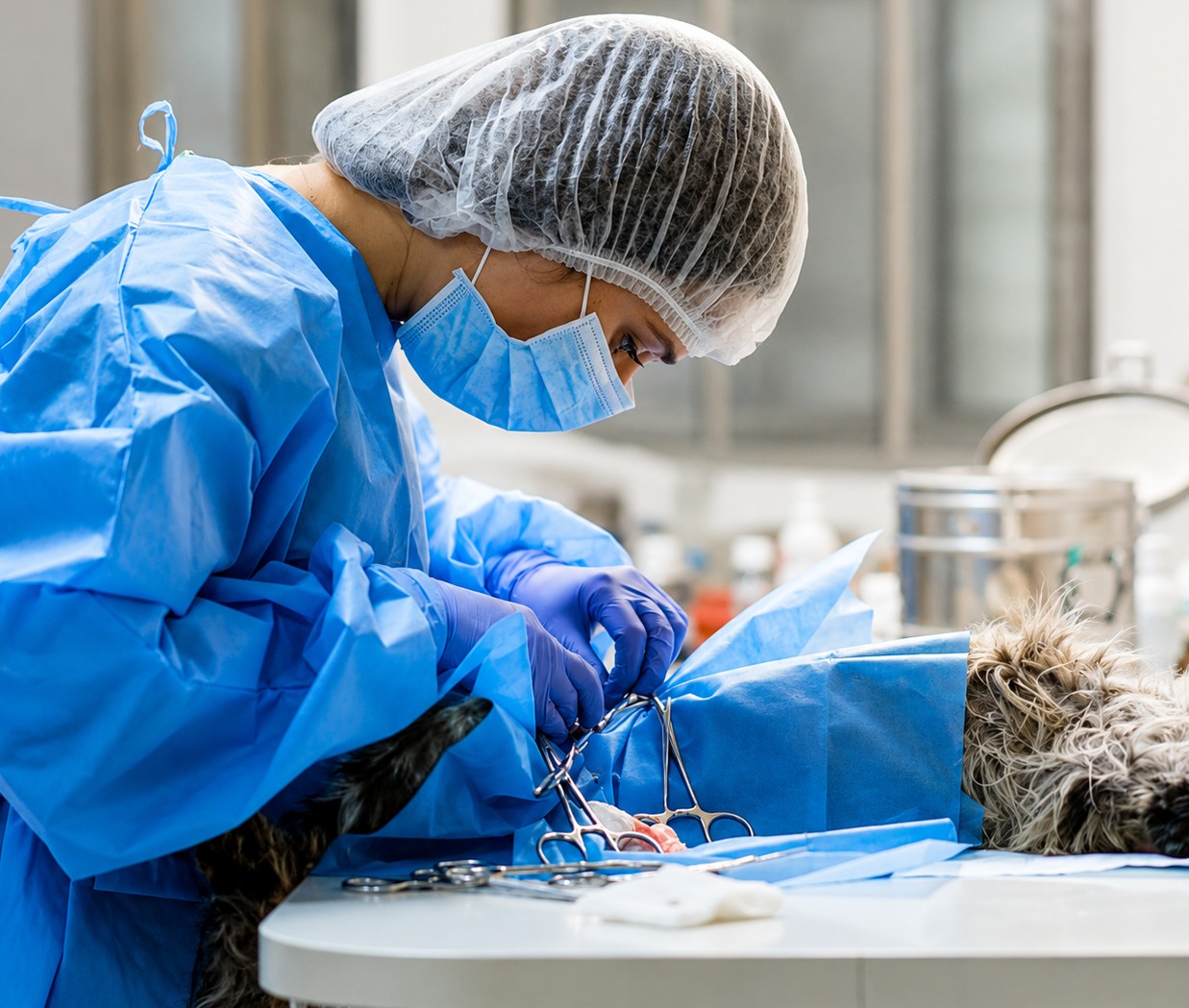
While PDS suture material degrades through hydrolysis in a relatively predictable manner compared to enzymatically degraded materials, several patient and environmental factors can influence the pace of this process. Understanding these variables is important for clinical interpretation of cases where PDS appears to be breaking down faster or slower than expected.
A significant inflammatory response at the surgical site, particularly one driven by bacterial infection, can accelerate the hydrolytic degradation of PDS. Elevated local temperature, changes in tissue pH associated with bacterial metabolism, and increased tissue fluid turnover all alter the hydrolytic environment around the suture. In infected wounds, PDS may lose tensile strength faster than the standard timeline predicts, which can have serious consequences if the tissue has not yet developed sufficient intrinsic strength to maintain the closure independently.
This is one of the reasons why preventing surgical site infection is so important in procedures that rely on PDS for prolonged structural support. If infection develops and accelerates suture degradation before the tissue has healed, the mechanical support the suture was selected to provide is no longer reliably present.
In tissues with very high vascularity and active metabolic turnover, the ready availability of water molecules and the dynamic tissue fluid environment may accelerate hydrolysis modestly. This effect is generally minor for PDS given its inherently slow degradation profile, but it is a relevant consideration in specific tissue environments.
Geriatric patients and those with significant systemic disease may process suture degradation products at a different rate than young, healthy animals. While PDS degradation itself is primarily a chemical process rather than a biological one, the elimination of breakdown products and the tissue response to ongoing hydrolysis are influenced by the patient’s overall metabolic function.
Larger diameter PDS sutures have a greater mass of polymer to degrade and therefore take longer to fully absorb than finer gauges of the same material. A size 0 PDS placed in the linea alba of a large breed dog will be physically present in tissue longer than a size 3-0 placed in the fascia of a small cat, even though both follow the same general degradation pattern.
In a small number of veterinary patients, PDS suture material does not follow its expected absorption trajectory. Signs suggesting abnormal degradation fall into two broad categories: breakdown that appears to be occurring too slowly, with the suture persisting beyond its expected absorption window; and breakdown that produces an unexpectedly pronounced tissue reaction, suggesting the degradation process is provoking a more significant inflammatory response than normal.
In thin-skinned patients or in areas where the subcutaneous tissue is minimal, an internal PDS closure may be palpable through the overlying skin as a linear or segmental firmness. This is generally normal within the first several months post-operatively. If this firmness persists beyond eight to ten months without reducing in intensity, this may suggest that absorption is proceeding more slowly than expected.
Persistent palpable suture material alone, in the absence of associated clinical signs, does not necessarily require intervention. However, it warrants monitoring and documentation so that its clinical significance can be assessed over time.
Post-operative imaging including ultrasonography, radiography, or advanced imaging that reveals distinct suture material at the implantation site beyond ten to twelve months post-operatively may suggest unusually slow absorption. As with palpable findings, the absence of associated clinical signs reduces the urgency of intervention, but the finding should be documented and monitored.
Although PDS suture material provokes significantly less tissue reaction than natural absorbable sutures, granuloma formation can occasionally occur with PDS, particularly during the active degradation phase when suture fragments are most abundant in the tissue. A granuloma presents as a firm, often non-painful nodule at or near the internal suture placement site. It may be palpable or only detectable on imaging depending on its depth.
Granulomas associated with PDS degradation are typically sterile and often resolve spontaneously as absorption progresses and the fragment burden decreases. If a granuloma persists, enlarges, or becomes associated with a draining tract, further investigation and potentially surgical management may be warranted.
A sinus tract emerging at the skin surface in the region of an internal PDS closure, producing persistent seropurulent discharge, indicates that the suture reaction has become pathological. This finding is uncommon with synthetic monofilament sutures such as PDS but can occur in immunocompromised patients, in the presence of concurrent infection, or when the suture material has been exposed to a particularly reactive tissue environment.
Management typically involves identifying the source of the tract through clinical examination and imaging, followed by surgical exploration and removal of the implicated suture material if spontaneous resolution does not occur.
Progressive swelling, warmth, or discomfort concentrated along the internal closure line beyond the expected early healing phase suggests an exaggerated tissue response to PDS degradation. This presentation is distinct from normal post-operative swelling in its progression rather than resolution over time.
In these cases, differentiating between a sterile exaggerated reaction to PDS degradation and an early surgical site infection is a clinical priority, as the management approaches differ significantly. Culture of any accessible discharge and assessment of systemic inflammatory markers may be helpful in making this distinction.
Certain patient populations are at higher risk of abnormal PDS breakdown and should be monitored more closely after procedures where PDS is used for internal closure.
Dogs receiving systemic immunosuppressive therapy, those with protein-losing conditions causing hypoproteinemia, dogs with severe concurrent disease, and dogs with a prior history of suture reactions all represent patient categories where PDS degradation may not follow the standard timeline. In these patients, more frequent post-operative check-ups and lower thresholds for further investigation of any unusual clinical findings are appropriate.
One of the most important practical applications of understanding PDS suture absorption time is using this knowledge to set appropriate client expectations at the time of discharge. Owners should be informed that the internal sutures used in their dog’s or cat’s surgery will take several months to fully dissolve, that some firmness along the closure line is normal and expected during this period, and that the body processes the suture material gradually without it needing to be removed.
This information prevents unnecessary concern and reduces the number of emergency contacts generated by owners who notice a firmness or palpable ridge along the incision site weeks after surgery and assume something has gone wrong.
For procedures using PDS in mechanically demanding locations such as abdominal wall closure or orthopedic soft tissue repair, a structured post-operative monitoring schedule supports early detection of any abnormal findings. An initial recheck at 10 to 14 days assesses early healing, a further recheck at four to six weeks confirms that the tissue is gaining appropriate strength, and a final assessment at three months can confirm that healing is complete and that no delayed suture reaction is developing.
When signs suggesting abnormal PDS breakdown are identified, the clinical response should be proportionate to the severity of the presentation. Mild sterile granuloma formation without clinical signs warrants monitoring. Progressive swelling, discharge, or sinus tract formation warrants investigation including imaging and potentially culture. Confirmed infection requires appropriate antimicrobial therapy and assessment of whether surgical exploration is needed.
The broader framework for recognizing and managing suture reactions in veterinary patients is covered in understanding suture reactions in dogs for better post-op recovery.
Ensuring that PDS degrades normally begins with appropriate clinical use from the outset. Placing PDS in tissues where its extended support duration is genuinely required, selecting the correct gauge for the patient and tissue, maintaining aseptic technique to prevent infection that could accelerate degradation, and avoiding placement in chronically inflamed or highly contaminated environments all support normal, predictable absorption.
For practitioners seeking a systematic approach to selecting PDS alongside other suture materials for specific tissue types, the suture selection guide for different tissue types in animals provides a comprehensive tissue-by-tissue reference.
Are PDS sutures dissolvable? Yes, completely and predictably, through hydrolysis over a timeline of six to eight months for full physical absorption. Is pds suture absorbable in the clinical sense that matters for surgical planning? Absolutely, with tensile strength support extending to six weeks and complete tissue elimination by eight months in most patients under normal conditions.
The clinical value of PDS suture material lies precisely in this extended, predictable absorption profile. Used in the right tissue, in the right patient, with the right technique, PDS provides the prolonged internal support that slower-healing tissues require while eventually degrading completely without a permanent foreign body remaining. Recognizing the signs when this process deviates from expectation, and responding appropriately, completes the clinical competency required to use this material effectively.
Strouden supplies high-quality PDS suture material and a full range of absorbable veterinary sutures to support reliable, predictable surgical outcomes. To explore our product range or discuss the right suture solutions for your practice, please contact us today.
A: Yes, PDS sutures are fully absorbable. They degrade through hydrolysis, a chemical process in which water molecules break down the polymer chains. Complete physical absorption typically occurs between 180 and 240 days post-implantation, making PDS one of the longest-absorbing synthetic absorbable sutures available.
A: PDS retains approximately 70 percent tensile strength at two weeks, 50 percent at four weeks, and around 25 percent at six weeks. Strength is largely lost by 8 to 10 weeks. Complete physical absorption of the suture mass occurs over 180 to 240 days in most patients.
A: Signs include a firm palpable suture line persisting beyond ten months, granuloma formation, progressive swelling or discomfort along the closure site, sinus tract formation with discharge, or imaging findings showing distinct suture material well beyond the expected absorption window.
A: Yes. Significant surgical site infection accelerates hydrolysis by altering local tissue pH and temperature. This can cause PDS to lose tensile strength faster than expected, potentially before the tissue has developed sufficient intrinsic strength, which may lead to wound dehiscence in severe cases.
A: Both are synthetic absorbable sutures degrading through hydrolysis, but their timelines differ significantly. Polyglactin 910 loses most tensile strength by three to four weeks and absorbs fully within 56 to 70 days. PDS retains strength for up to six weeks and takes 180 to 240 days for complete absorption.