
Contents
Among the many synthetic absorbable suture materials available to veterinary professionals today, polyglycolic acid suture occupies a well-established and widely trusted position. Known by its abbreviation PGA, this braided synthetic absorbable material has been in clinical use for decades and continues to be a reliable choice for a broad range of internal tissue closure applications across veterinary specialties. For practitioners who encounter PGA suture regularly and want a thorough understanding of its composition, absorption mechanism, and clinical behavior, this article provides a complete reference.
PGA stands for polyglycolic acid, a synthetic polymer produced from glycolic acid monomers arranged into long polymer chains. Polyglycolic acid was among the first fully synthetic absorbable suture materials developed for medical use, introduced in the early 1970s as an alternative to natural absorbable materials such as catgut that offered more predictable and consistent absorption behavior.
The manufacturing process for PGA absorbable suture involves extruding the polymer into fine filaments, which are then braided together to form the suture strand. This braided construction is the standard form in which PGA suture is supplied for veterinary use. The braided structure gives the material its characteristic handling properties, including excellent pliability, good knot security, and ease of manipulation in the surgical field.
Some PGA sutures are supplied with a coating, typically a combination of calcium stearate and other lubricating agents, which further enhances handling by reducing tissue drag during placement without significantly altering the absorption profile. Coated and uncoated versions of polyglycolic acid suture are both available, and the choice between them is generally based on surgeon preference and specific application requirements.
PGA suture has a distinctive appearance compared to monofilament absorbable materials. Its braided construction creates a slightly textured surface that is visible at close inspection, and the suture has a softer, more pliable feel than monofilament alternatives such as polydioxanone or poliglecaprone 25. This pliability is one of the key practical advantages of PGA in clinical use and contributes to its continued popularity alongside newer materials.
The color of PGA suture is typically violet or undyed beige depending on the manufacturer, with the violet dye added to improve visibility in tissue during placement. Undyed versions are available for applications where dye presence is undesirable.
Polyglycolic acid suture is absorbed through hydrolysis, the same chemical degradation process that breaks down other synthetic absorbable sutures including polyglactin 910, polydioxanone, and poliglecaprone 25. In hydrolysis, water molecules present in tissue fluid penetrate the suture material and cleave the ester bonds linking the glycolic acid monomers together. This progressive bond cleavage reduces the polymer chains to shorter and shorter fragments until the material is fully broken down into glycolic acid, which enters normal metabolic pathways and is eliminated from the body.
Because PGA degrades through hydrolysis rather than enzymatic breakdown, its absorption rate is considerably more consistent and predictable than that of natural absorbable sutures such as chromic catgut. Enzymatic degradation depends on the local concentration of degrading enzymes, which varies between tissue types, patients, and clinical conditions. Hydrolysis proceeds at a rate primarily determined by the availability of water molecules and the local chemical environment, making it inherently more uniform across different patients and tissue locations.
This predictability is one of the primary reasons synthetic absorbable sutures including PGA became the standard replacement for natural absorbable materials in most veterinary surgical applications.
The glycolic acid produced by PGA hydrolysis enters the body’s normal biochemical pathways. It is converted in the liver to glycine, which is then metabolized further through pathways that ultimately produce carbon dioxide and water as end products. This metabolic pathway is well established and produces no accumulation of toxic byproducts under normal physiological conditions, which is why PGA absorbable suture is considered safe for use in internal tissue environments across all commonly encountered veterinary patient species.
The polyglycolic acid suture absorption time follows a well-documented pattern that veterinary professionals can use to match PGA to applications where its support duration is appropriate.
PGA retains approximately 65 percent of its original tensile strength at two weeks post-implantation. By three weeks, this has reduced to approximately 35 percent. At four weeks, only a small residual fraction of tensile strength remains. In most implantation environments, meaningful tensile strength has been largely lost by three to four weeks after placement.
This strength retention timeline is significantly shorter than that of polydioxanone, which retains 50 percent of its strength at four weeks, and makes PGA most appropriate for tissues that heal relatively quickly and where prolonged mechanical support beyond three to four weeks is not required.
The physical mass of PGA suture material takes longer to be fully eliminated from tissue than the loss of tensile strength would suggest. Complete absorption of polyglycolic acid suture typically occurs within 60 to 90 days post-implantation in most veterinary patients under normal conditions.
During this period the suture becomes progressively fragmented and reduced in volume as hydrolysis continues. The remaining material is increasingly low-mass and biologically inert as degradation advances, and the tissue response to it diminishes correspondingly. By 90 days, the vast majority of PGA suture material has been eliminated from the implantation site in healthy patients.
Suture Material |
Strength Lost By |
Complete Absorption |
Construction |
| Poliglecaprone 25 | 2 to 3 weeks | 90 to 120 days | Monofilament |
| Polyglycolic acid (PGA) | 3 to 4 weeks | 60 to 90 days | Braided |
| Polyglactin 910 | 3 to 4 weeks | 56 to 70 days | Braided |
| Chromic catgut | 10 to 14 days | 60 to 90 days | Twisted natural |
| Polydioxanone (PDS) | 8 to 10 weeks | 180 to 240 days | Monofilament |
This comparison positions PGA clearly within the short-to-medium-term support category, alongside polyglactin 910, and distinguishes it from longer-acting materials like polydioxanone that are selected when tissue healing is expected to be slow.
The local tissue environment has a meaningful influence on PGA absorption rate. In highly vascularized tissues with active metabolic turnover, the ready availability of water and the dynamic fluid environment can accelerate hydrolysis, resulting in somewhat faster absorption than the standard timeline predicts. Conversely, in poorly vascularized or relatively avascular tissues, absorption may proceed more slowly.
For practical purposes, the 60 to 90 day complete absorption window provides a reliable planning reference for most clinical applications, with the understanding that individual variation is possible and generally modest.
Significant infection or inflammation at the surgical site can accelerate PGA degradation by altering local pH and increasing tissue fluid turnover. In infected wounds, PGA may lose tensile strength faster than the three to four week standard timeline, potentially leaving the wound without suture support before adequate tissue strength has developed. This is a relevant consideration in any case where post-operative infection develops in a wound closed with PGA.
Patient age, nutritional status, and systemic health influence the metabolic processing of PGA degradation products and the tissue’s capacity to manage the ongoing hydrolysis process. Young, healthy animals generally process PGA absorption efficiently. Patients with severe systemic disease or significant metabolic compromise may show variability in absorption behavior, though this effect is more pronounced with enzymatically degraded natural sutures than with hydrolytically degraded synthetic materials like PGA.
PGA absorbable suture is widely used for subcutaneous tissue closure in veterinary patients of all species. Its excellent handling characteristics, good knot security, and appropriate short-to-medium-term support duration make it practical and reliable for eliminating dead space and reducing tension on the overlying skin closure. In clean elective surgical procedures, the modest infection risk associated with braided construction is generally acceptable in the relatively low-contamination subcutaneous environment.
The superior knot security of braided PGA suture makes it a reliable choice for vessel ligation and organ pedicle ties during procedures such as ovariohysterectomy, castration, and organ resection. The knot must hold securely through the early post-operative period to maintain hemostasis, and the braided construction of PGA provides the friction-based knot security necessary for this application. Its consistent absorption within 60 to 90 days aligns well with the healing timeline of ligated pedicles.
Oral mucosal tissues heal rapidly due to the rich vascularity of the oral cavity. PGA suture is frequently used for gingival flap closure, oral mucosal repair, and palatal procedures in veterinary patients. The confined working space and the need for reliable knot security in a moist, enzyme-rich environment favor braided construction, and PGA’s 60 to 90 day absorption timeline is more than sufficient for the relatively fast healing of oral tissues.
PGA is used in some gastrointestinal surgical applications, though the preference for monofilament sutures in this environment is well established due to bacterial wicking concerns. In clean gastrointestinal cases or for outer layer closures where contamination risk is lower, PGA can be appropriate. For inner layer closures directly contacting luminal contents in contaminated cases, monofilament absorbable sutures are the standard recommendation. This distinction is discussed in what vets need to know about braided vs monofilament suture.
For urogenital procedures including bladder closure, uterine repair, and associated pedicle ligation, PGA is used selectively. For pedicle ligation in the body cavity during ovariohysterectomy, braided PGA provides reliable hemostatic security. For mucosal surfaces in direct contact with urine, monofilament absorbable sutures are preferred to minimize the risk of suture calculi formation around braided material.
Across a broad range of soft tissue procedures including tumor excision, wound reconstruction, and exploratory laparotomy closure, PGA suture is used for muscle and fascial layer approximation in cases where extended support is not required. Its three to four week strength retention is appropriate for most muscle closures in healthy patients, and its handling ease makes it a practical choice for general soft tissue work.
A comprehensive guide to PGA applications across veterinary specialties is available in common polyglycolic acid suture uses across different vet specialties.
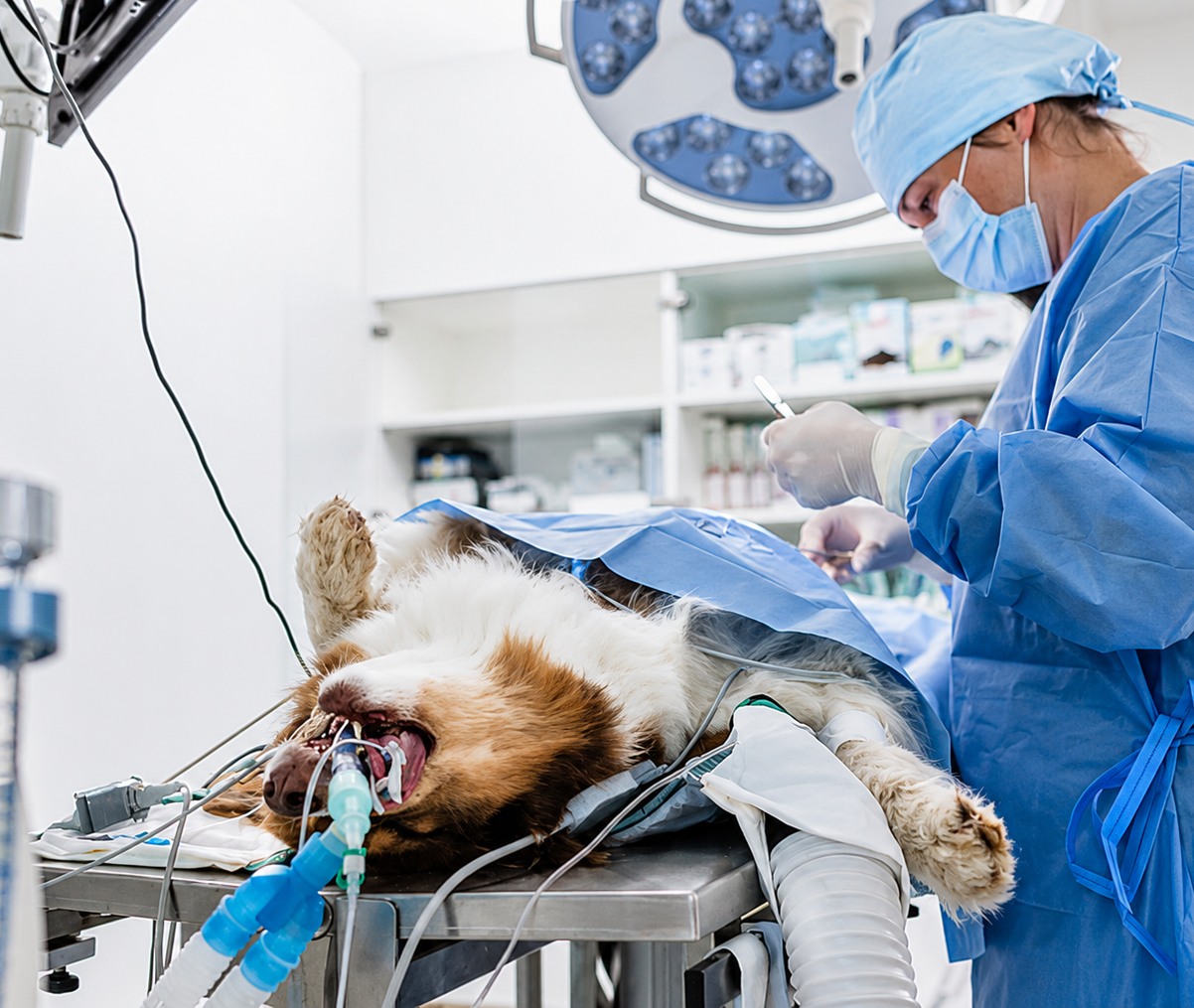
The braided construction of PGA absorbable suture gives it excellent pliability and knot security that practitioners consistently cite as practical advantages. It handles more easily than monofilament sutures, requires fewer knot throws to achieve reliable security, and behaves predictably during placement in a variety of tissue environments. For procedures where surgical efficiency matters or where less experienced practitioners are performing closure, these handling characteristics have genuine value.
The hydrolytic degradation of PGA proceeds consistently across the vast majority of patients and tissue environments, giving surgeons reliable planning reference points for support duration and absorption timeline. This predictability reduces the uncertainty that can accompany use of enzymatically degraded natural sutures, where individual variation in degradation rate is more pronounced.
PGA is well tolerated by most animal tissue types. While its braided construction provokes a modestly greater inflammatory response than monofilament synthetic alternatives, the reaction is generally mild in healthy patients and resolves as the suture absorbs. In the clean tissue environments where PGA is most appropriate, this level of tissue response rarely produces clinical complications.
The braided structure of PGA creates microscopic spaces where bacteria can harbor and form biofilm, sheltered from immune defenses and antibiotic penetration. In contaminated wounds, gastrointestinal mucosal closures, and other high bacterial load environments, monofilament absorbable sutures are strongly preferred. For these applications, polydioxanone or poliglecaprone 25 provide the monofilament construction necessary for infection risk minimization.
PGA’s three to four week tensile strength retention makes it unsuitable for slowly healing tissues such as fascia, abdominal wall, and orthopedic soft tissue repairs where support for six or more weeks is clinically necessary. For these applications, polydioxanone is the appropriate choice. Selecting PGA in a slow-healing tissue environment risks losing suture support before the tissue has developed adequate intrinsic strength, increasing the risk of wound breakdown.
Application |
Recommended Material |
Reason |
| Abdominal wall closure | PDS | Extended support required |
| Subcutaneous closure | PGA or polyglactin 910 | Short support sufficient, knot security valued |
| Intestinal anastomosis | PDS or poliglecaprone 25 | Monofilament required for infection control |
| Organ pedicle ligation | PGA or polyglactin 910 | Knot security critical, short support adequate |
| Fascial repair | PDS | Six-week support required |
| Oral mucosal closure | PGA or polyglactin 910 | Rapid healing, braided handling practical |
PGA suture should be stored in cool, dry conditions within the manufacturer-specified temperature and humidity range. Excess heat or moisture can accelerate hydrolysis of the suture material within its packaging, reducing tensile strength before the suture is placed in the patient. Packaging integrity should be confirmed by visual inspection before opening, and any packet showing moisture staining, seal compromise, or physical damage should be discarded.
As a synthetic absorbable suture with a defined shelf life, PGA stock should be managed using a first-in, first-out rotation system to prevent expiry dates from being missed. Guidance on veterinary surgical supply management is available in how to maintain medical equipment for long-term reliability.
Understanding what is pga suture in both compositional and clinical terms gives veterinary professionals the foundation to use this material effectively and selectively. Polyglycolic acid suture is a braided synthetic absorbable material that degrades through hydrolysis, losing tensile strength within three to four weeks and achieving complete tissue absorption within 60 to 90 days. These properties make it well suited to subcutaneous closure, ligature applications, oral surgery, and general soft tissue approximation in clean surgical fields where its handling advantages can be fully realized without the infection risk concerns that apply in contaminated environments.
By matching PGA to the applications where its properties genuinely serve the patient’s healing needs, and by selecting alternative materials such as polydioxanone where extended support or monofilament construction is required, veterinary professionals can use polyglycolic acid suture as the reliable and clinically valuable tool it has proven to be across decades of veterinary surgical practice.
Strouden supplies high-quality PGA absorbable suture and a comprehensive range of veterinary surgical sutures to support consistent, well-informed surgical practice. To explore our full product range or discuss the best suture options for your practice, please contact us today.
A: PGA stands for polyglycolic acid, a synthetic absorbable suture made from braided glycolic acid polymer filaments. It degrades through hydrolysis, breaking down into glycolic acid that is metabolized and eliminated. It is one of the most widely used absorbable suture materials in veterinary surgery.
A: PGA suture loses most tensile strength by three to four weeks post-implantation. Complete physical absorption of the suture mass typically occurs within 60 to 90 days in most veterinary patients under normal tissue conditions and without significant infection or inflammation at the implantation site.
A: PGA is most commonly used for subcutaneous tissue closure, organ pedicle ligation, oral mucosal repair, and general soft tissue approximation in clean surgical fields. Its excellent knot security and handling ease make it practical for ligature applications and procedures requiring reliable braided construction.
A: PGA is not recommended for contaminated wounds because its braided construction creates capillary spaces where bacteria can harbor and form biofilm. In contaminated environments, monofilament absorbable sutures such as polydioxanone or poliglecaprone 25 are preferred for their superior resistance to bacterial wicking.
A: PGA loses tensile strength by three to four weeks and absorbs fully within 60 to 90 days, making it suited to fast-healing tissues. PDS retains strength for up to six weeks and absorbs over 180 to 240 days, making it appropriate for slowly healing tissues such as fascia and abdominal wall.