
Contents
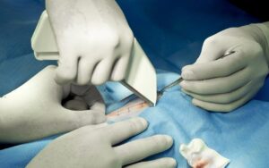
In veterinary surgery, polydioxanone sutures have become a trusted choice due to their excellent tensile strength, smooth monofilament design, and reliable absorption. Widely used across various tissue types and specialties, these sutures offer predictable degradation within the body. For veterinarians, understanding the absorption time of polydioxanone sutures is key to optimizing their effectiveness and ensuring the best outcomes in surgery.
This article covers the science behind how polydioxanone breaks down in animal tissue, the factors that influence its absorption timeline, the clinical scenarios where it performs best, and how it compares to other medical sutures in the absorbable category.
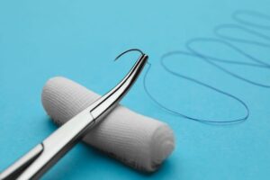
Polydioxanone, commonly referred to as PDS sutures, is a synthetic absorbable monofilament suture produced from a polyester polymer. It was developed specifically to address the need for a suture material that could maintain mechanical support over a longer period than standard absorbable options such as polyglactin 910 or polyglycolic acid, while still degrading completely within the body.
PDS sutures are widely used in veterinary medicine for procedures involving slowly healing tissues, high-tension closures, and body cavities where prolonged internal support is necessary. Their monofilament construction means they pass smoothly through tissue with minimal drag and do not harbor bacteria in the way braided sutures can.
For veterinary professionals looking to understand how PDS fits within the broader landscape of suture options, the guide to the different types of veterinary surgical sutures provides a useful overview.
The absorption of PDS sutures occurs through a process called hydrolysis. Hydrolysis involves the gradual breakdown of the polymer chains that make up the suture material when they come into contact with water molecules present in body tissues. This is a chemical reaction rather than an enzymatic one, which means it proceeds at a relatively consistent and predictable rate regardless of the specific tissue environment.
The products of polydioxanone hydrolysis are metabolized safely by the body and eliminated without significant accumulation. This metabolic pathway contributes to the low tissue reactivity associated with PDS sutures, as the body does not mount a significant immune response to the breakdown products.
This hydrolytic mechanism contrasts with the absorption of natural sutures such as chromic catgut, which rely on enzymatic breakdown. Enzymatic degradation is more variable because it depends on the local concentration of degrading enzymes, which can fluctuate with infection, inflammation, or individual patient differences. The more controlled nature of hydrolysis is one reason synthetic absorbable sutures, including PDS, are preferred in modern veterinary practice for most clinical applications.
The polydioxanone suture absorption time follows a well-documented pattern that makes it a reliable choice for surgical planning.
PDS sutures retain their mechanical strength for significantly longer than most other absorbable medical sutures. The approximate timeline is as follows.
At two weeks post-implantation, PDS sutures retain approximately 70 percent of their original tensile strength. At four weeks, roughly 50 percent of strength remains. By six weeks, strength has reduced to approximately 25 percent. Complete loss of mechanical strength typically occurs by around 8 to 10 weeks.
This extended strength retention is the defining clinical advantage of PDS sutures. It means the suture continues to provide meaningful mechanical support well into the proliferative and early remodeling phases of wound healing, which is particularly valuable in tissues where collagen synthesis and maturation are slow.
While tensile strength loss is largely complete by 8 to 10 weeks, the physical mass of the suture material continues to be present in the tissue for a longer period. Complete absorption of PDS sutures typically occurs between 180 and 240 days, which translates to approximately six to eight months post-implantation.
This does not cause clinical concern in the vast majority of cases. The suture material becomes progressively fragmented and reduced in mass as hydrolysis continues, and the remaining material is biologically inert and well tolerated by surrounding tissues. Veterinary professionals should be aware of this timeline when advising clients about what to expect following surgery, particularly if post-operative imaging is performed and suture remnants are visible.
For comparison with other absorbable suture timelines, how long does absorbable sutures last in veterinary procedures provides a comprehensive reference across multiple material types.
While the polydioxanone suture absorption time is more predictable than that of enzymatically degraded materials, several factors can still influence the pace of breakdown.
Highly vascularized tissues with active metabolic turnover may experience slightly faster hydrolysis because water is more readily available and tissue fluid exchange is more dynamic. Conversely, poorly vascularized tissues may slow the hydrolytic process modestly.
A significant inflammatory response at the surgical site can accelerate suture degradation. Elevated local temperatures, changes in tissue pH, and increased enzymatic activity associated with infection can all contribute to faster breakdown of the polymer chains. This is an important consideration when PDS sutures are placed in potentially contaminated environments.
Larger diameter sutures take longer to fully absorb because there is a greater mass of polymer to break down. A size 0 PDS suture will persist in tissue longer than a size 3-0, even though the tensile strength retention pattern follows a similar curve proportionally.
Age, nutritional status, and systemic health can all influence the rate of tissue metabolism and therefore the pace of suture hydrolysis. Young, healthy animals with robust metabolic function may process suture materials slightly differently than geriatric or debilitated patients. These individual variations are generally modest with PDS sutures compared to enzymatically degraded materials, but they remain clinically relevant in complex cases.
The extended polydioxanone suture absorption time makes PDS particularly well suited to several specific clinical contexts.
Fascia is a dense, relatively avascular connective tissue that heals slowly and experiences significant mechanical loading during the post-operative period. The abdominal wall in particular must withstand the internal pressures generated by respiration, defecation, and movement. PDS sutures are a frequent choice for linea alba and fascial closure because their prolonged tensile strength retention supports the wound through the full critical healing period.
In orthopedic surgery, soft tissue structures including joint capsules, periosteum, and muscle fascia require support through an extended healing period. PDS sutures are used in these applications because they bridge the gap between the immediate post-operative period and the point at which the repaired tissue gains sufficient intrinsic strength. Their role in joint repair procedures is discussed in how orthopedic sutures are used in veterinary joint repair procedures.
For bladder closures, uterine repairs, and similar urogenital procedures, PDS sutures offer the combination of monofilament construction and prolonged support that these tissues require. The smooth surface reduces bacterial wicking in environments exposed to urine, and the extended absorption timeline accommodates the variable healing rates seen in urogenital tissues.
Young animals undergoing surgery benefit from absorbable sutures that eliminate the need for a stressful removal procedure. PDS sutures provide sufficient support for developing tissues while eventually degrading completely. Their monofilament structure also reduces the risk of irritation in smaller, more delicate tissue planes.
In large or heavily muscled patients, the forces exerted on surgical closures are proportionally greater than in small animals. PDS sutures provide the extended mechanical support needed to prevent wound breakdown during the longer healing periods associated with high body mass and activity levels.
A practical surgical suture guide would be incomplete without a comparison between PDS and other commonly used absorbable materials.
Polyglactin 910 (Vicryl): This braided absorbable suture retains approximately 50 percent of its tensile strength at two weeks and loses most of its strength by three to four weeks. Complete absorption occurs within 56 to 70 days. It is appropriate for tissues that heal relatively quickly and where excellent knot security is important, but it does not provide the prolonged support of PDS.
Polyglycolic Acid (PGA): Similar in profile to polyglactin 910, PGA retains strength for approximately two to three weeks with complete absorption by 60 to 90 days. It is a reliable multifilament absorbable suture but, like polyglactin, is better suited to shorter-term support requirements. Its clinical applications are detailed in common polyglycolic acid suture uses across different vet specialties.
Poliglecaprone 25 (Monocryl): This monofilament absorbable suture loses its strength relatively quickly, retaining approximately 50 percent at one week and 20 to 30 percent at two weeks. Complete absorption occurs within 90 to 120 days. It is well suited to subcuticular skin closure and rapidly healing tissues but is not appropriate for slow-healing or high-tension applications.
Chromic Catgut: This natural absorbable suture retains strength for approximately 10 to 14 days and is completely absorbed within 60 to 90 days through enzymatic breakdown. Its variable absorption profile and higher tissue reactivity make it less predictable than synthetic options. Further detail on its use is available in exploring chromic catgut suture uses in animal soft tissue repair.
Among these options, PDS stands out for applications requiring support beyond the four-week mark. No other commonly used absorbable suture provides the same combination of monofilament construction and extended strength retention.
PDS sutures are generally well regarded for their handling characteristics, though they require some adaptation compared to braided materials. As a monofilament, PDS has inherent memory, meaning it tends to spring back toward its original coiled shape. This requires additional knot throws to achieve secure knot tying, and practitioners new to the material benefit from deliberate practice to develop reliable technique.
Despite this, the advantages of monofilament construction, particularly reduced tissue drag and lower infection risk, generally outweigh the handling challenges. Modern formulations of PDS have improved pliability compared to earlier versions, making the material easier to work with in a range of tissue environments.
Proper storage of PDS sutures is important to maintain their performance. Sutures exposed to excessive heat, humidity, or light may experience premature degradation of the packaging or the suture material itself. Practices can review best standards for maintaining their surgical supply inventory in the guide to buying wholesale medical supplies.
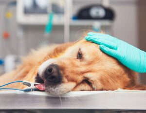
Because PDS sutures are used primarily in internal tissues, post-operative monitoring focuses on the surgical site rather than the suture material directly. Clients should be advised to monitor for signs of wound complication including swelling, discharge, heat, or behavioral changes suggesting pain. Regular follow-up examinations allow the veterinary team to confirm that healing is progressing appropriately and that the suture is providing the intended support.
In cases where imaging is performed after surgery, PDS sutures may be visible as a linear density on ultrasound or radiograph for several months post-implantation. This is normal and reflects the extended physical absorption timeline rather than a complication.
Understanding polydioxanone suture absorption time gives veterinary professionals a clear framework for when and why to select PDS sutures over other available absorbable medical sutures. With tensile strength retention extending to six to eight weeks and complete physical absorption occurring over six to eight months, PDS occupies a unique position among absorbable materials, bridging the gap between short-term absorbable sutures and permanent non-absorbable options. Its monofilament construction adds further clinical value through reduced tissue reactivity and bacterial resistance.
Strouden supplies high-quality PDS sutures and a full range of veterinary surgical materials to support consistent, reliable outcomes across every surgical application. To find out more about our product range or to discuss the best suture solutions for your practice, please contact us today.
A: PDS sutures retain approximately 70 percent of tensile strength at two weeks, 50 percent at four weeks, and around 25 percent at six weeks. This extended retention makes them well suited for slow-healing tissues and high-tension closures in veterinary patients.
A: Complete physical absorption of polydioxanone sutures typically occurs between 180 and 240 days post-implantation. While strength is largely lost by 8 to 10 weeks, the suture material continues to degrade gradually through hydrolysis over the following months.
A: Yes, polydioxanone is a common choice for fascial and abdominal wall closure because its prolonged tensile strength retention supports the wound through the slow healing process of dense connective tissue. Its monofilament construction also reduces infection risk in body cavity closures.
A: Yes, significant infection or inflammation can accelerate hydrolysis by altering local tissue conditions including pH and temperature. This may cause PDS sutures to lose strength faster than expected, which is one reason contaminated wounds require careful management alongside appropriate suture selection.
A: Polyglactin 910 loses most of its tensile strength by three to four weeks and absorbs fully within 56 to 70 days. PDS retains strength significantly longer and absorbs over six to eight months. PDS is preferred for slow-healing tissues while polyglactin 910 suits faster-healing internal closures requiring good knot security.