
Contents
When veterinary professionals discuss suture selection, the conversation most often centers on the suture material itself. The choice between absorbable and non-absorbable materials, braided and monofilament constructions, and specific polymers such as polydioxanone or polyglactin 910 receives significant attention in clinical education and practice. However, the veterinary suture needle attached to that material is an equally important component of the closure system, and its selection has direct consequences for tissue trauma, passage ease, closure accuracy, and healing outcomes.
Choosing the right needle for a given tissue type, anatomical location, and procedure requires understanding needle geometry, point design, body curvature, and suture needle length. This article provides a comprehensive guide to veterinary suture needle selection, explaining the key characteristics of available needle types and how those characteristics translate into clinical performance across the range of procedures encountered in veterinary surgical practice.
A suture needle is not merely a delivery mechanism for the suture material. It is a precision tissue instrument that determines how much damage occurs with each pass through the tissue, how easily the suture is directed through the intended tissue plane, and how accurately wound edges can be approximated. A needle that is poorly matched to the tissue being sutured increases trauma with every pass, makes precise suture placement more difficult, and ultimately contributes to a less favorable healing environment.
The relationship between needle selection and tissue trauma is particularly important in delicate or highly vascularized tissues such as intestinal mucosa, vascular structures, and ophthalmic tissue. In these locations, using a needle with inappropriate point geometry or an oversized body creates damage that accumulates with each successive pass and can meaningfully affect local healing capacity.
For a broader understanding of how needle choice fits within the complete wound closure decision, understanding veterinary surgical wound closure techniques for better recovery provides a thorough contextual reference.
Even in more robust tissues such as skin and muscle, the cumulative effect of repeated needle passes with an inappropriately matched instrument contributes to unnecessary inflammation and slower recovery. Proper needle selection is therefore a meaningful contributor to the overall quality of surgical wound closure, alongside the material and pattern choices that typically receive more attention.
Every suture needle consists of three distinct anatomical regions, each of which contributes specific functional characteristics.
The point is the tissue-penetrating tip of the needle. Its geometry determines how the needle enters tissue, how much force is required for penetration, and what type of tissue channel the needle creates as it passes through. Point design is the most clinically significant aspect of needle selection for most applications because it determines the extent of tissue trauma with each pass.
The body is the main curved section of the needle that the needle holder grasps and controls during suture placement. Body geometry determines the needle’s curvature, its cross-sectional profile, and its resistance to bending under the forces applied during suturing. The body cross-section varies between needle types, with round-bodied, cutting, and reverse-cutting designs each presenting different cross-sectional geometries that influence how the needle behaves in tissue.
The swage is the attachment point where the suture material is permanently joined to the needle. Most modern suture needles used with commercial veterinary surgical sutures are swaged, meaning the suture is embedded into a hollow at the needle’s tail end during manufacturing. This swaged attachment creates a smooth, uninterrupted transition between needle and suture that minimizes the tissue channel created compared to older eyed needles through which suture was threaded, doubling the material passing through each needle hole.
Taper point needles have a smooth, gradually tapering body that comes to a sharp point without cutting edges along the body. As the needle passes through tissue, it spreads the tissue fibers apart rather than cutting through them, creating a channel through which the suture passes without excising any tissue. When the needle is withdrawn, the elastic tissue fibers close back around the suture, producing a snug fit that minimizes leakage of fluid along the suture line.
Taper point needles are the standard choice for soft, easily penetrable tissues including intestine, peritoneum, subcutaneous fat, muscle, and vessel walls. Their tissue-spreading action makes them the preferred choice in any tissue where maintaining structural integrity around the suture channel is important. They are used extensively in gastrointestinal surgery, abdominal closure, and vascular applications.
For guidance on suture material selection to accompany taper point needles in these tissue types, the suture selection guide for different tissue types in animals provides a comprehensive tissue-by-tissue reference.
Because taper point needles do not cut tissue, they are not appropriate for dense or tough tissues such as skin or fascia, which resist penetration without a cutting edge. Attempting to use a taper point needle in dense tissue requires excessive force that increases the risk of needle bending or breaking.
Cutting needles have two opposing cutting edges running along the inner curvature of the needle body, with a third cutting edge at the point. This triangular cross-sectional profile creates a sharp instrument that cuts through tissue with each pass. The cutting edges are located on the inside of the needle’s curve, meaning the needle cuts toward the wound edge as it passes through tissue.
Cutting needles are used in tough, dense tissues that resist penetration by taper point needles. Skin is the most common application for cutting needles in veterinary practice, along with oral mucosa, ligaments, and other fibrous or dense tissue structures. The sharp cutting action allows these resistant tissues to be penetrated with lower force and more controlled placement.
The inside cutting edge position does have a clinical limitation. Because the cutting edge faces the wound margin, there is a tendency for suture tension to enlarge the needle hole toward the wound edge, which can contribute to suture pull-through in tissues under high tension. This has led to the development of the reverse cutting needle as the preferred alternative in many applications.
The reverse cutting needle has the same triangular cross-sectional profile as a standard cutting needle, but the third cutting edge is located on the outer curvature of the needle body rather than the inner. This repositions the flat edge of the triangular profile toward the wound margin, providing a flat inner surface against which the suture sits rather than a cutting edge that could enlarge the needle hole under tension.
Reverse cutting needles are the most widely used needle type in veterinary practice for skin closure. Their cutting action allows reliable penetration of the dermis and epidermis while the flat inner surface reduces the risk of suture pull-through under tension. They are also used for subcutaneous tissue closure, tendon repair, and other applications in relatively dense tissue where reliable penetration and suture security are required.
Understanding which suture materials pair best with reverse cutting needles for skin closure is covered in how to choose the best sutures for skin closure in vet practice.
The reverse cutting design has largely replaced the standard cutting needle in contemporary veterinary suture needle selection for most skin and soft tissue applications.
The tapercut needle combines the penetration advantage of a cutting point with the tissue-spreading body of a taper needle. The tip section has a short cutting portion that initiates penetration through tough tissue, while the body transitions to a round taper profile that spreads rather than cuts as it passes through. This hybrid design is engineered to penetrate dense tissue at the point while minimizing the tissue channel created by the body.
Tapercut needles are used in specific applications where tissue is too dense for a pure taper point but where the full cutting body of a reverse cutting needle would create a larger-than-necessary tissue channel. Cardiovascular surgery involving calcified vessel walls, tendon repair in chronically inflamed tissue, and orthopedic soft tissue closures are among the typical applications.
For procedures involving orthopedic soft tissue repair, the role of suture material selection alongside appropriate needle choice is discussed in how orthopedic sutures are used in veterinary joint repair procedures.
Blunt point needles have a rounded, non-cutting tip that cannot penetrate intact tissue under normal suturing forces. They are designed for friable, fragile tissues where a conventional sharp point carries a significant risk of inadvertent puncture of adjacent structures.
Blunt point needles are used primarily in liver and kidney parenchymal tissue, where the organ’s friable structure means a sharp needle point can easily create unintended lacerations in adjacent tissue. In veterinary practice, blunt point needles are encountered most commonly in hepatic or renal procedures and in some soft tissue applications where organ parenchyma must be sutured.
Spatula needles have a flat, spatula-like body with cutting edges on the top and bottom of the flat profile rather than on the sides. This geometry allows the needle to pass through tissue in a precise horizontal plane, splitting tissue layers apart without creating a cylindrical channel. The flat profile minimizes vertical deflection of the needle during passage.
Spatula needles are used almost exclusively in ophthalmic surgery. The precise horizontal passage they provide is essential when suturing the thin, multilayered tissues of the cornea and sclera, where vertical deflection could cause penetration into unintended tissue planes. Veterinary ophthalmic surgeons rely on spatula needles for corneal laceration repair and keratoplasty procedures.
Needle body curvature determines how much rotation of the needle holder is required to complete a full needle pass through tissue, how accessible different anatomical locations are with a given needle, and how easily the needle can be directed through confined surgical spaces. Curvature is expressed as a fraction of a circle, with common options including one quarter circle, three eighths circle, half circle, and five eighths circle.
For context on how curvature selection integrates with tissue layer closure decisions, how long does absorbable sutures last in veterinary procedures provides relevant background on the tissue environments where different needle curvatures are most commonly applied.
Quarter circle needles require minimal wrist rotation to complete a pass. They are used in ophthalmology and in very superficial tissue planes where a small arc of needle travel is appropriate.
The three eighths circle is the most widely used needle curvature in general veterinary surgery. It strikes a practical balance between accessibility and maneuverability, requiring a moderate arc of wrist rotation that most surgeons find intuitive and controllable. It is the standard curvature for skin closure, subcutaneous closure, and most soft tissue procedures.
Half circle needles require a full 180-degree wrist rotation to complete a pass. This greater arc makes them useful in confined or deep spaces where the needle must travel a longer arc to reach and exit the tissue in a controlled manner. They are commonly used in body cavity closures including abdominal wall repair and in gastrointestinal anastomoses.
Five eighths circle needles complete the greatest arc and are designed for very confined spaces where a straight or minimally curved needle would not be navigable. Urological procedures and deep pelvic surgery represent typical applications.
Straight needles are used in accessible tissue locations where the needle can be passed and retrieved without the need for a curved path. Skin closure in easily accessible locations and some subcuticular applications may use straight needles, though their use is considerably less common than curved designs in most veterinary surgical contexts.
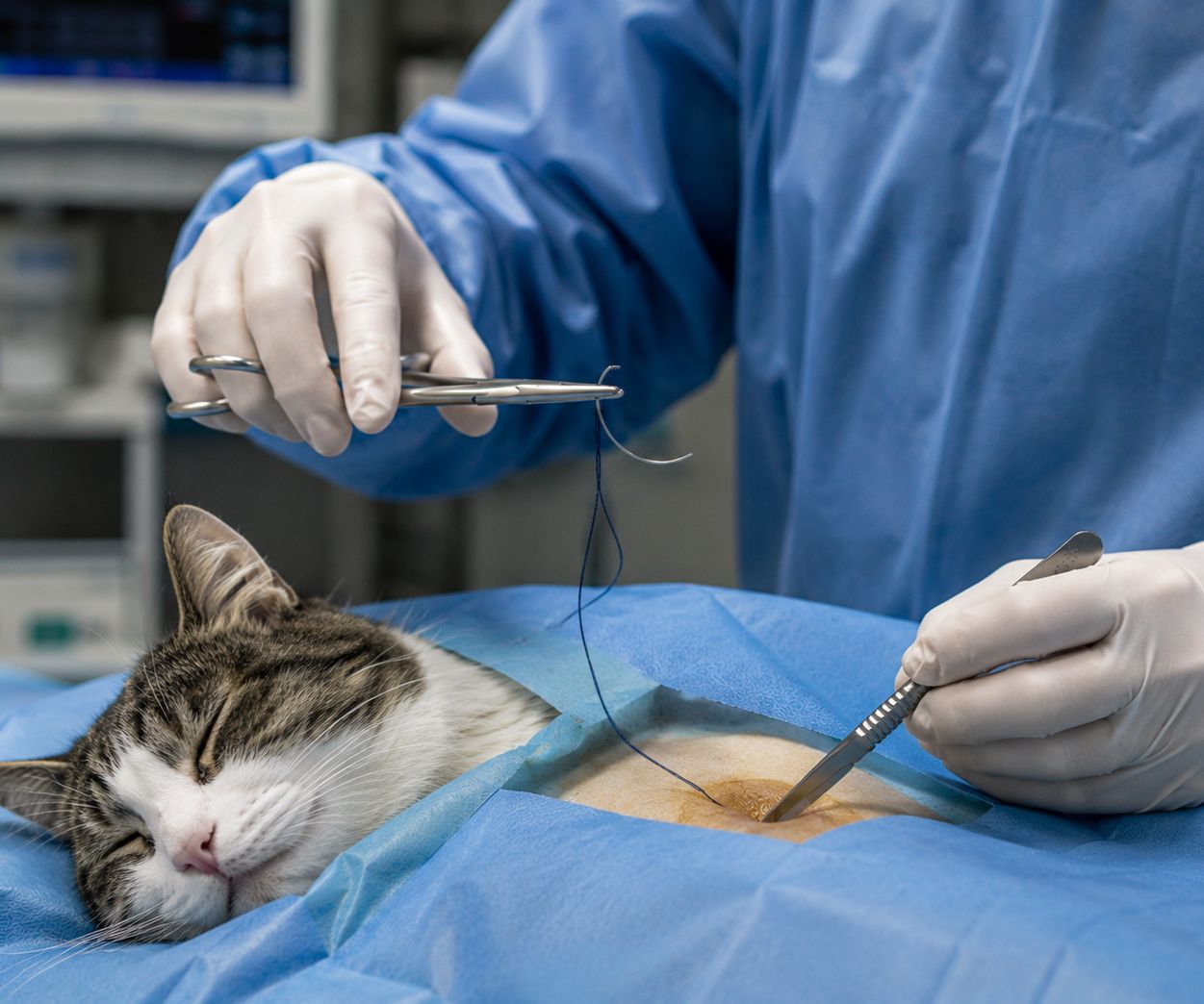
Suture needle length determines how far the needle travels through tissue with each pass and how much tissue is incorporated in each suture bite. Appropriate suture needle length selection ensures that the suture bite depth is matched to the tissue thickness being closed, that the needle can be reliably retrieved after passing through the tissue, and that the needle can be controlled accurately throughout its arc of travel.
A needle that is too short will not allow adequate tissue bites, resulting in shallow sutures that may pull through under tension. A needle that is too long is more difficult to control and increases the risk of the needle tip contacting adjacent structures during its arc of travel.
For skin closure in small animals, shorter needle lengths in the range of 16 to 26 millimeters are generally appropriate. For body wall closure, longer needles in the range of 26 to 36 millimeters allow adequate tissue bites through the full thickness of the abdominal wall. For ophthalmic procedures, very short needles of 5 to 13 millimeters provide the precision required in small anatomical structures.
Large animal applications require longer needles to accommodate greater tissue thickness, with skin and muscle closure in horses and cattle commonly using needles in the 40 to 60 millimeter range. Appropriate gauge selection for suture material accompanying these needle lengths is discussed in how to choose suture size for optimal healing and vet care.
Suture needle length and body curvature interact to determine the practical arc of needle travel during placement. A longer needle with a three eighths circle curvature travels a larger physical arc than a shorter needle with the same curvature fraction. Understanding this interaction allows surgeons to select needle dimensions that match the spatial constraints of the specific anatomical working environment.
Needle gauge refers to the thickness of the needle wire, which determines its bending strength and resistance to deformation under suturing forces. Heavy-gauge needles resist bending in tough tissues but create larger channels with each pass. Fine-gauge needles produce smaller tissue holes but are more prone to bending under high resistance.
Needle gauge should match the tissue resistance expected during the procedure. Skin closure in large breed dogs requires a heavier gauge needle than corneal closure in a cat, and the needle supplied with the suture should reflect this. Most commercial veterinary surgical sutures are supplied with needles matched to the gauge and application of the suture material, which simplifies selection in routine cases.
Procedure |
Recommended Point |
Recommended Curvature |
Notes |
| Skin closure | Reverse cutting | 3/8 circle | Standard choice across species |
| Subcutaneous closure | Reverse cutting or taper | 3/8 circle | Taper if tissue is soft |
| Abdominal wall closure | Taper | 1/2 circle | Tissue spreading preferred |
| Gastrointestinal anastomosis | Taper | 1/2 circle | Minimizes mucosal trauma |
| Vascular repair | Taper | 3/8 or 1/2 circle | Fine gauge, precise placement |
| Oral mucosal closure | Reverse cutting or taper | 3/8 circle | Depends on tissue density |
| Corneal repair | Spatula | 1/4 circle | Ophthalmic specific |
| Tendon repair | Tapercut or reverse cutting | 3/8 circle | Depends on tissue condition |
| Liver or kidney parenchyma | Blunt | 1/2 circle | Prevents inadvertent puncture |
| Large animal skin closure | Reverse cutting | 3/8 circle | Heavier gauge, longer length |
Commercial veterinary surgical sutures are supplied as complete needle and suture systems in which the needle has been selected to match the suture material gauge, the intended tissue application, and the expected performance requirements. Using the needle supplied with the suture material as a complete matched system ensures that the needle diameter is appropriate for the suture gauge and that the swage attachment is optimized for the specific material.
Attempting to reuse needles or to attach different suture materials to needles not designed for them risks swage failure, needle deformation, and compromised closure integrity. Sourcing sutures from reliable suppliers who provide consistently matched needle and suture systems is therefore an important component of ensuring consistent surgical outcomes.
For guidance on selecting absorbable suture materials that pair appropriately with different needle types across tissue applications, key differences in absorbable vs non-absorbable sutures every veterinarian should know provides a useful reference.
The suture material used in a procedure should inform needle point selection as well as the tissue type. Monofilament sutures such as polydioxanone or nylon pass through tissue most smoothly when paired with appropriate needle geometries that minimize friction during passage. Braided sutures have more surface friction regardless of needle type but benefit from reverse cutting or taper designs that create clean channels rather than ragged ones. The structural implications of different suture constructions are covered in what vets need to know about braided vs monofilament suture.
Suture needles are precision instruments and their performance degrades with use even within a single procedure. A needle that has been passed through tissue multiple times, that has contacted a hard surface such as bone or an instrument, or that has been improperly grasped by the needle holder can develop a blunted point, a barb along the body, or a bent tip that significantly increases tissue trauma with each subsequent pass.
Surgeons should assess needle condition during lengthy procedures and replace the needle and suture assembly if any degradation in penetration ease or directional control is noted. Continuing to use a compromised needle increases tissue trauma and can compromise the final quality of the closure.
The needle holder should grasp the needle at the junction of the middle and rear thirds of the body, avoiding the swage region where suture attachment could be compromised by instrument pressure. Grasping the needle too close to the point risks tip damage, while grasping at the swage risks disrupting the suture attachment. Consistent correct placement preserves needle integrity across multiple passes during a single closure.
The broader context of maintaining surgical instrument standards in veterinary practice is discussed in how to maintain medical equipment for long-term reliability.
Selecting the right veterinary suture needle is an exercise in matching instrument geometry to tissue biology. Point design determines how tissue is entered and what channel is created. Body curvature determines how the needle travels through the surgical space. Suture needle length determines how much tissue is incorporated in each bite. Together these characteristics define how the needle performs in a given tissue and anatomical context.
Developing a systematic approach to veterinary needles selection, grounded in an understanding of these properties and their clinical implications, elevates surgical technique and contributes to better healing outcomes for every patient. The role of supply quality in supporting consistent needle and suture performance is discussed in the importance of medical supplies in building trust.
Strouden supplies a comprehensive range of veterinary surgical sutures with matched needle systems across all standard needle types, curvatures, and suture needle lengths to support precise, tissue-appropriate wound closure. To explore our full product range or discuss the right suture and needle systems for your practice, please contact us today.
A: The reverse cutting needle with a three eighths circle curvature is the most widely used needle for skin closure in veterinary practice. Its cutting action allows reliable penetration of dense dermis, while the flat inner edge reduces suture pull-through risk compared to standard cutting needle designs.
A: Suture needle length determines the depth and width of each tissue bite. A needle too short produces shallow sutures prone to pull-through, while an overly long needle is harder to control and risks contacting adjacent structures. Matching length to tissue thickness supports accurate, reliable closure.
A: Taper point needles are used in soft, easily penetrable tissues including intestine, peritoneum, subcutaneous fat, and vessel walls. They spread tissue fibers rather than cutting them, producing a snug channel around the suture that minimizes fluid leakage and reduces tissue trauma in delicate internal structures.
A: Spatula needles are used for corneal and scleral repair in veterinary ophthalmic surgery. Their flat profile allows passage through the precise horizontal plane of corneal tissue without vertical deflection, which is essential for accurate repair of the thin, multilayered structures of the eye.
A: Needle curvature determines how much wrist rotation is required to complete each pass and how accessible confined anatomical spaces are with a given needle. A three eighths circle suits most general procedures, while a half circle is preferred for body cavity closures where a greater arc of travel through confined tissue is required.