
Contents
Surgical success in veterinary medicine hinges significantly on the meticulous art and science of surgical wound closure. The careful selection and application of appropriate surgical wound closure techniques are paramount for promoting optimal tissue integrity, minimizing complications, and ensuring rapid, comfortable recovery for animal patients. This comprehensive guide dives into the various methods and materials used, emphasizing their impact on healing outcomes.
Wound closure is one of the most important aspects of any surgical procedure, directly influencing how efficiently animals recover and how well tissues reconnect. Proper closure not only facilitates apposition of tissue layers but also provides immediate tensile strength, protecting the surgical site from external contamination and mechanical stress. The goal is to restore anatomical and functional integrity, leading to minimal scarring and a strong, resilient repair.
Veterinary clinicians understand that effective wound management extends beyond the incision itself. It encompasses the entire process from initial assessment to postoperative care. The choice of surgical sutures types and closure methods must be tailored to the specific tissue, the degree of tension, and the expected healing environment. This critical decision-making process underscores the expertise required in every surgical moment.
To effectively select appropriate surgical wound closure techniques, a foundational understanding of the wound healing process is essential. Wound healing is a complex biological cascade, traditionally divided into four overlapping phases:
Immediately after injury, hemostasis, or the cessation of bleeding, occurs through vasoconstriction and clot formation. This is quickly followed by the inflammatory phase, where immune cells migrate to the wound site to clear debris and pathogens. This phase is characterized by redness, swelling, heat, and pain, all critical for initiating the reparative process.
During the proliferative phase, granulation tissue forms, characterized by new blood vessels (angiogenesis), fibroblasts producing collagen, and epithelial cells migrating to re-establish the skin barrier. This phase is crucial for filling the wound defect and providing a scaffold for subsequent tissue remodeling.
The final phase, remodeling, involves the maturation of collagen fibers, increasing tensile strength of the healing tissue. This process can continue for months to years, gradually strengthening the repair. The type of tissue and the closure technique can significantly influence the efficiency and completeness of this stage.
Choosing the correct material for surgical wound closure is as vital as the technique itself. Several factors dictate this choice, including the tissue type, the wound’s location, the desired duration of support, and potential for infection. Strouden offers a comprehensive range of high-quality veterinary surgical supplies to meet these diverse needs.
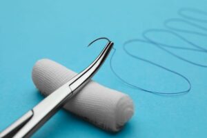
Absorbable sutures are designed to lose their tensile strength and break down within the body over time, eliminating the need for removal. Their degradation occurs typically through hydrolysis or enzymatic breakdown. These are ideal for internal tissues where prolonged support is not required or when suture removal would be difficult.
Common types include:
Polyglicolic Acid (PGA): A synthetic braided multifilament suture known for its high initial tensile strength and predictable absorption profile.
Polydioxanone (PDS) / Poliglecaprone (PGCL): Synthetic monofilament sutures offering smooth passage through tissue and extended tensile strength retention, suitable for slower-healing tissues.
Learn more about how long dissolvable stitches last after vet surgery for insights into their absorption characteristics.
Non-absorbable sutures retain their tensile strength indefinitely and are used when permanent tissue support is required, such as in cardiovascular, neurological, or orthopedic procedures, or for skin closure where removal is feasible. They elicit minimal tissue reaction.
Common types include:
For a detailed comparison, explore our guide on monofilament suture vs. multifilament for clinical use.
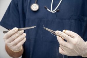
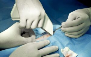
Mastery of various suturing techniques is a hallmark of skilled veterinary professionals. The chosen technique depends on the tissue being approximated, the desired aesthetic outcome, and the need for tension relief.
Each stitch is tied individually, providing high security. If one suture fails, the others remain intact. This technique is often used for skin closure or in tissues under tension.
Executed as a single, continuous strand of suture material. This method is faster but if one part of the suture breaks, the entire line can unravel. It’s often used for fascia, subcutaneous tissue, or to provide an air-tight or water-tight seal.
Beyond basic interrupted and continuous patterns, several specialized techniques address unique surgical challenges:
Proper application of these techniques ensures optimal apposition and minimizes opportunities for complications, reinforcing positive healing outcomes.
While sutures are fundamental, modern veterinary practice incorporates a range of other surgical wound closure techniques that offer unique advantages in specific scenarios.
Disposable skin staplers offer a fast and efficient method for skin closure, particularly in long incisions or in situations requiring rapid closure due to patient instability. They are useful in areas not subject to excessive movement. Staples provide good wound edge eversion and are generally easy to remove post-operatively. However, proper technique is crucial to avoid excessive pressure on wound edges, which can lead to localized necrosis. For veterinary procedures, understanding the practices to remove surgical staples from animal wounds ensures optimal patient comfort and healing.
Veterinary skin glue, or topical skin adhesive, polymerizes upon contact with tissue, forming a strong, flexible bond. It is excellent for closing small, superficial incisions or lacerations with minimal tension, particularly in areas difficult to bandage or in fractious animals where rapid closure is beneficial. Skin glue can significantly reduce surgical time and eliminate the need for suture removal. The key benefits of vet skin glue for faster healing in pets make it a valuable tool in many clinical settings.
While less common in primary veterinary surgical closure compared to human medicine, adhesive strips can play a role in reinforcing primary closures or for very superficial wounds. They provide light support and approximation, often preventing the need for deep sutures.
Optimal healing outcomes are not solely dependent on the chosen surgical wound closure techniques but on a holistic approach to patient care.
Gentle tissue handling throughout the surgical procedure is crucial. Excessive crushing, tearing, or stretching of tissues can impede blood supply and delay healing. Sharp dissection with appropriate instruments minimizes collateral damage.
Maintaining strict asepsis is non-negotiable in surgery. Contamination can lead to infection, the most common and devastating complication in wound healing. How to sterilize medical equipment is a critical procedure that prevents surgical site infections and ensures patient safety. Strouden understands the importance of medical supplies in building trust and consistently provides high-quality, sterile products.
Effective hemostasis prevents hematoma formation, which can act as a culture medium for bacteria and mechanically separate tissue layers, delaying healing. Ligature use, electrocautery, and pressure contribute to controlling hemorrhage.
Dead space, an area within a wound devoid of tissue approximation, can fill with fluid (seroma) or blood (hematoma). This creates an ideal environment for bacterial growth and can delay healing. Obliterating dead space through appropriate suturing or placement of drains is essential.
Proper postoperative care, including pain management, bandage changes, and activity restriction, significantly impacts recovery. Clients must be educated on how to care for their pet’s incision site and recognize signs of complications. Ensuring that veterinary practices have access to and know how to maintain medical equipment for long-term functionality is also key to providing consistent, high-quality care.
Veterinary surgeons frequently encounter challenges that demand careful consideration and adaptable surgical wound closure techniques.
Tension across a wound edge can lead to dehiscence (wound breakdown) or excessive scarring. Strategies to manage tension include releasing incisions, tension-relieving suture patterns, and undermining of the skin to allow for greater mobility. For large defects, skin grafts or flaps may be necessary.
In these cases, primary closure may be contraindicated. Delayed primary closure, secondary closure, or healing by second intention (allowing the wound to heal naturally by granulation and contraction) might be more appropriate. Extensive debridement and appropriate antimicrobial therapy are critical.
Some tissues, such as those that are inflamed or devitalized, are delicate and prone to tearing. Using larger bite sizes, reinforcing sutures, or opting for less traumatic suture materials like monofilaments can help.
The field of veterinary medicine is continually evolving, with innovations contributing to improved surgical wound closure techniques and patient outcomes. From advanced suture materials with antimicrobial coatings to sophisticated stapling devices designed for specific anatomical regions, these advancements provide veterinary professionals with more tools to deliver superior care. Keep abreast of the latest for optimal patient care; explore our guide to buying wholesale medical supplies to ensure your clinic is well-equipped.
By staying informed about the latest research and product developments, veterinarians can continue to refine their practice, choosing the most effective and least invasive methods for each unique patient. This dedication to continuous improvement directly translates into better healing outcomes and enhanced quality of life for animals.
In conclusion, mastering surgical wound closure techniques is foundational to achieving superior recovery in veterinary patients. The thoughtful selection of suture materials, the skillful application of various suturing methods, and the judicious use of alternative closure options like staples and skin glue, all contribute to successful surgical interventions. At Strouden, we are dedicated to providing veterinary professionals with the high-quality supplies necessary for these critical procedures. We empower you with the tools to ensure optimal healing outcomes and build trust with your clients through exceptional patient care. To learn more about our comprehensive range of veterinary surgical solutions, please do not hesitate to contact us. Together, we can promote faster, safer, and more comfortable recoveries for all animal patients.
A: The primary types of surgical wound closure techniques include suturing, stapling, adhesive tapes, and tissue adhesives (surgical glue). The choice depends on the wound’s depth, location, tension, and desired cosmetic outcome.
A: Sutures work by physically bringing the edges of a wound together, allowing the body’s natural healing process to bridge the gap. They can be absorbable, dissolving over time, or non-absorbable, requiring removal.
A: Surgical stapling is often preferred for closing long incisions, in areas with high tension, or when speed is critical, such as in emergency surgeries. It can also be beneficial in certain internal organ anastomoses.
A: No, surgical glue (tissue adhesive) is typically used for small, superficial, low-tension wounds and often in conjunction with deeper sutures. It’s not suitable for deep, jagged, or infected wounds, or areas under significant stress.
A: Primary wound closure involves directly closing the wound edges immediately after surgery, aiming for minimal scarring. Secondary wound closure leaves the wound open to heal from the bottom up, often used for contaminated or infected wounds.